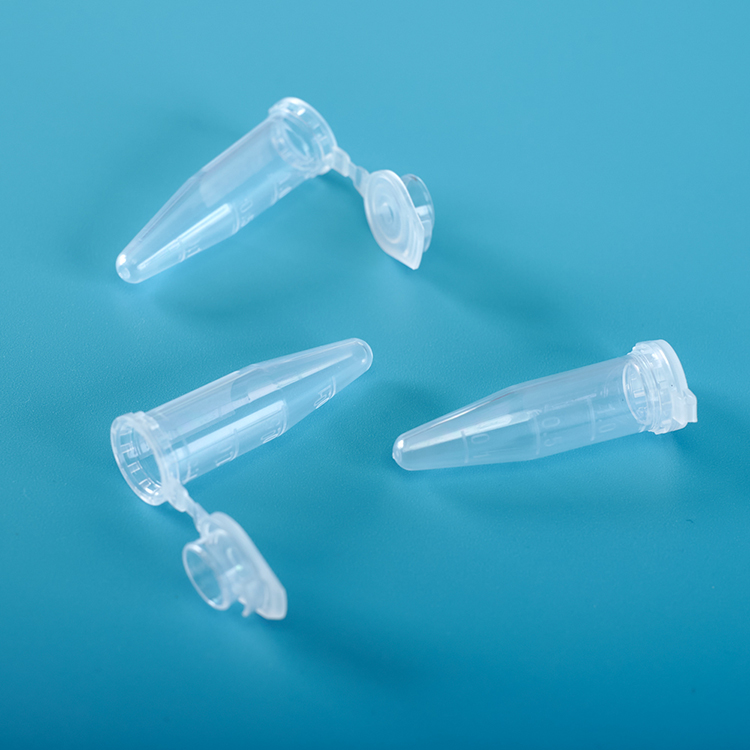
Centrifuge Tubes
Centrifuge tubes are used in laboratory centrifuges, machines that spin samples in order to separate solids out of liquid chemical solutions. The centrifuge tubes can be made of glass or plastic, and resemble miniature test tubes with tapered tips. They are mainly used in series test like the centrifuge of nucleic acid
50 ml Centrifuge Tubes,15 ml Centrifuge Tubes,5ml Centrifuge Tubes,Centrifuge Tube Holder,Micro centrifuge Tubes Yong Yue Medical Technology(Kunshan) Co.,Ltd , https://www.yonyue.com
FITC-N=C=S+N-H2-Protein→FITC-NS-CN-H2-Protein The fluorescent antibody is usually labeled by the Marsshall (1958) method. It can also be based on conditions using the Chadwick et al. marker method or Clark et al. (1963). Marking method.
1.Marsshall method
(1) Materials: Antibody globulin solution, 0.5 mol/L (pH 9.0) carbonate buffer, sterile saline, fluorescein isothiocyanate, 1% thimerosal water solution, 50 ml beaker, 4°C refrigerator, Magnetic stirrer, dialysis bag, glass rod, 0.01 mol/L PBS pH 7.2 or 3.0, and the like.
(2) Method and Step 1 Preparation of Antibody: Take a suitable amount of globulin solution of known concentration in a beaker, add human physiological saline and carbonate buffer, so that the final immunoglobulin concentration is 20 mg/ml, carbonate The buffer volume is 1/10 of the total volume. Mix well and place on a magnetic stirrer at a rate of 5 to 10 minutes.
2 Preparation of Fluorescein: According to the total amount of protein to be labeled, add 0.01mg of fluorochrome per mg of immunoglobulin, and accurately weigh the required fluorescein isothiocyanate powder with an analytical balance. The amount of immunoglobulin and fluorescein can also be calculated by the following formula, and the amount of buffer to be added can also be calculated.
a. Protein solution: content Amg/m1; volume Bml.
b. Total protein amount (AXB) = Crag.
cC/20 ~ C/10 = Dmg (if the protein content is less than 20 mg/ml, use C/10; if higher than 20 mg/ml, use C/20).
d. The amount of fluorescein FITC: (1/50 to 2/100) XC=Emg.
e. å·³ 0.5 mol/L (pH 9.5) carbonate buffer D/10 = Fml.
f. PBS amount D-(B+F) = Gml.
Note: A is protein content, mg/ml; B is protein solution volume; C is total protein, mg; D is constant, mg; , ml; G is the volume of PBS, ml.
3 Binding (or labeling): Gradually add the fluorochrome weighed into the globulin solution while stirring to avoid sticking the fluorescein to the wall of the flask (approximately within 5 to 10 minutes). After the addition is complete, continue the dark light mixing. 12h or so. The protein solution should be kept at about 4°C during the binding process, so it is necessary to transfer the beaker and the stirrer to a 4°C refrigerator.
4 Dialysis: After the binding is completed, the labeled globulin solution is centrifuged (2500 r/min) for 20 minutes, a small amount of precipitate is removed, placed in a dialysis bag, placed in a beaker, and dialyzed with pH 8.0 buffered saline (0- 4~C) overnight.
5 column: take the overnight dialysis marker, Sephadex G-25 or G-50 column Sephadex gel, isolated free fluorescein, collect the labeled fluorescent antibody for identification. Eluent: 0.01 mol/L phosphate buffer (pH 7.2); Filtration amount: 12 ml labeled global protein solution (no dialysis before filtration); Collection amount: 20 ml (diluted 1.7 times or so).
2.Chadwick method
(1) Reagents and materials: Antibody globulin solution, fluorescein isothiocyanate, 3% sodium carbonate aqueous solution, 0.01 mol/L pH8.0 PBS, 1% thimerosal, centrifuge and centrifuge tube, beaker (25 ml) stirrer, no Bacteria straws, sterile straws and capillary drops, beakers (500 ml), dialysis bags, etc.
(2) Method and Step 1 Antibody preparation: pH 4.0 with o~4~C. 0 Phosphate buffered saline The globulin solution was diluted to a concentration of 30 ~ 40 mg/ml, placed in a 25 ml beaker, and placed in an ice bath.
2 Preparation of fluorescent pigment: Calculate the amount of fluorescein 0.01rug per mg of immunoglobulin, weigh the desired fluorescein, and dissolve it with 3% sodium carbonate aqueous solution.
3 Mix the prepared antibody with the fluorescent dye solution in equal amounts, stir well, and combine it in the o~4~C refrigerator (preferably on a magnetic stirrer) for 18~24h.
4 dialysis and column chromatography: method with the Marsshall method.
3. Improved method
(1) Reagents and Materials In a 10.01 mol/L (pH 7.2) PBS formulation, the calibration pH was set to 7.2. NaCi18g, Na2HP041.15g, dissolved in 2000ml distilled water 20.5mol/L (pH9.0) carbonate buffer, and 0.5ml/L NazCOs (5.3%) 10ml was added to 0.5ml/L NaHC03 (4.2%) 90ml and mixed. , calibration pH to 9.0.
A 33% aqueous solution of sodium carbonate was prepared by dissolving L5g anhydrous sodium carbonate in 50 ml of sterile distilled water.
4 other reagents and materials l% sodium azide, centrifuges and Centrifuge Tubes, beakers (25m1), stirrers, sterile pipettes and capillary drops, beaker (500m1) dialysis bags.
(2) Method and Step 1 High titer anti-human globulin rabbit immune serum, isolated globulin, diluted with saline (0.15 mol/L NaCl) and buffer (0.5 mol/L NaHC03-Na2C03, pH 9.0) Contains lOmg protein, the buffer is 10% of the total, cooling to 4 °C.
2 Add isothiocyanate (FITC) fluorescein [protein: fluorescein = (50-80) mg/mg] at 0-4°C.
Stir under electromagnetic stirring for 12 to 14 hours.
3The labeled globulin is then precipitated with semi-saturated ammonium sulfate to remove unbound fluorescein, and then dialyzed against buffered saline to remove ammonium sulfate (tested with Nessler's reagent until the saline dialyzed overnight has no ammonia ions and fluorochrome). .
4 The prepared fluorescent antibody plus azide sodium o.01%, subpackaged in lml ampoules, or freeze-dried, stored in the refrigerator (4 °C) can be used more than six months, a 20 ~ C save up to 2 years the above.
4. Dialysis Marking
This method is suitable for the fluorescein labeling of a small number of antibodies, and has a simple labeling and less non-specific staining.
(1) Reagents and materials: Reagents and materials with the modified method.
(2) Method and Step 1 Dilutions of the immunoglobulin to be labeled with 1% concentration in 0.025 mol/L carbonate buffer, pH 9.0, into a dialysis bag.
2 Using the same buffer, mix the FITC with a 0.1 mg/ml solution. At 10 times the volume of the 10 mg/ml globulin solution, fill the FITC diluent in a cylindrical container and immerse the dialysis tubing in the FITC solution.
3 The top of the container is tightly closed, stir bar is placed at the bottom, and the mark is dialyzed for 24 h under electromagnetic stirring at 4~C. The labeling solution in the dialysis bag was removed and gel filtration was immediately performed with Sephadex G50 to remove free fluorescein, which was then dispensed and stored at 4°C. 
FITC fluorescein-labeled antibody procedure
When FITC is reacted with an antibody protein in an alkaline solution, the amino group of lysine on the protein is mainly combined with the thiocarbamide of fluorescein to form a FITC-protein conjugate, that is, a fluorescent antibody or a fluorescent conjugate. . There are 86 lysine residues in an IgG molecule, which can generally bind 15-20 at most. An IgG molecule can bind 2-8 molecules of FITC. The reaction formula is as follows: